

Optical HREM Micro
Optical High-Resolution Episcopic Microscopy MICRO System for 3D Morphological Imaging
High-Resolution 3D Imaging with Micro
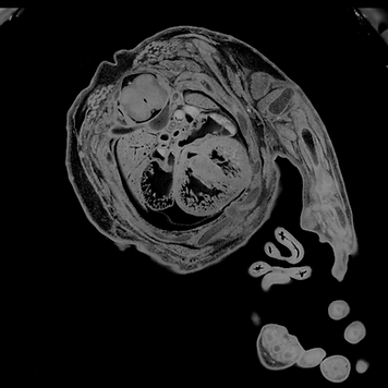
Optical HREM Micro is a high-resolution imaging system designed to generate 3D volumes from dense biological and agricultural samples. It captures precise 2D images and reconstructs them into a complete volumetric dataset, revealing detailed morphological structures across entire samples.
The system delivers simplified yet informative structural outputs, making it ideal for developmental biology, pathology and plant or agricultural research. Optical HREM Micro bridges the gap between traditional histology and 3D imaging, offering whole-sample insight with microscopic resolution.

Key Advantages of the Micro System
Designed for Morphological Analysis
Ideal for quantifying organ shape, tissue architecture, and structural development in embryology or agriscience studies.
Streamlined Workflow for Larger Samples
Easy-to-learn interface enables routine 3D imaging without requiring specialized training perfect for core facilities or academic labs.
Reliable Sectioning for Dense Samples
Provides uniform and precise sectioning across embryos, plant tissue and bone, even in optically dense material.
Interoperable Image Exports
Data outputs are fully compatible with FIJI/ImageJ, Amira, and Imaris for 3D reconstruction, segmentation, and volume rendering.
Whole-Sample 3D Imaging
Generates high-resolution 3D reconstructions of entire embryos, organoids, or tissue samples using serial block-face imaging.
Compatible with Plant and Animal Tissues
Used in imaging plant root systems, seed morphology, fixed mouse embryos, and other complex biological structures.

Use Cases for Whole-Sample 3D Imaging with Micro
-
3D Phenotyping in agricultural studies allow for visualisation of barley, seeds, chicken embryos in high contrast despite the density
-
Morphological analysis in developmental biology such as whole embryos, chick embryos and organoids to assess organ development, malformations or gene knockout effects
-
Whole-mount imaging in histopathology or toxicology in samples such as mouse organs zebrafish and drosophila
-
Tissue structure evaluation in educational labs
Micro for Streamlined HREM Imaging
Micro HREM is ideal for individuals or HREM experimentation allowing for full 3D imaging of structures.
-
Single channel, single shot imaging at 20 megapixels
-
Capable of 1 block, multiple samples per section (in the same shot)
-
Resolution depends on sample size, not on desired values, still capable of achieving 5-0.5-micron lateral.
OHREM Micro Specifications
Resolution
-
Variable axial resolution from 1-8 microns.
-
Lateral resolution is variable based on samples size 5 micron(FOV 25mm) - ~0.5 micron(FOV 1mm).
Imaging Time
-
~ 7 Seconds/Section
-
~39 Minutes/mm
Sample Size Capacity
-
1 Block, multiple samples in one block
-
Max block size 25mm, recommended 15mm x 12mm x 10mm
Image Output
-
File types: jpg, jpeg, tif, tiff, png
-
20 Megapixels Digital Resolution
-
2D Image stacks for use in 3D volume packages (Dragonfly etc).
Imaging Depth
-
Max imaging depth: 25mm
-
Typical volume stack: 2000-3000 sections @ 3 microns
Options
-
Dual/Multi fluorescence upgrade
-
Small XY motorisation
Contact our High-Resolution Episcopic Microscopy (HREM) Experts
Request a quote or a meeting to discuss the Optical HREM Micro instrument using the form.
Phone: +44(0) 1462633500